
Research Symposium -2024
Musculoskeletal Research Symposium
Friday, April 26, 2024
The Center for Biomechanical Engineering Research and the Delaware Center for Musculoskeletal Research will hold a joint Research Symposium at the North Atrium at STAR Campus. Our goal is to show case the exciting research performed by faculty, students, and clinicians at University of Delaware and surrounding hospitals (Nemours Children’s Hospital and Christiana Care), spanning from basic, translational, to clinical studies of biomechanical injury and repair processes and musculoskeletal disorders.
Keynote Speaker: Dr. Karl Lewis
Title: Fluorescent imaging advances for the study of osteocyte mechanotransduction mechanisms

Dr. Karl Lewis is an assistant professor in the Meinig School of Biomedical Engineering at Cornell University. Prior to starting his lab at Cornell, he completed his PhD training with Prof. Mitch Schaffler at The City College of New York, and a post-doctoral fellowship with Prof. Alex Robling at the Indiana University School of Medicine. The Lewis Lab is working on 3 main focus areas; namely investigation of bone mechanobiology, expanding non-linear microscopy approaches in bone tissue, and establishing the use of novel fluorescent probes for imaging cellular dynamics in vivo.
For more information, please see Dr. Karl Lewis’ page.
Abstract: Prof. Lewis and his group work to better understand the interplay of mechanical inputs and biological outcomes in bone. In service of this goal, they develop novel intravital imaging techniques for studying osteocyte mechanotransduction and mechanobiology in vivo. These approaches leverage fluorescent signal to observe cellular/molecular level events and allow for experiments that keep critical endocrine and tissue crosstalk pathways intact. As such, they offer powerful insights into cellular behavior in health and disease. In this presentation, Prof. Lewis will share new approaches and subsequent findings related to osteocyte mechanoregulation and endocytosis.
Program – subject to change
| 8:45 | Welcome & Introductory Remarks | DCMR Director: Dawn Elliott CBER Director: X. Lucas Lu |
| 9:00 | Keynote Lecture Title: Fluorescent imaging advances for the study of osteocyte mechanotransduction mechanisms |
Karl Lewis, PhD |
| 10:00 |
Faculty Talk 1 Title: SUPERLUBRICITY! Reconsidering Our Knowledge of Articular Cartilage Tribology and Function. |
Chris Price |
| 10: 15 | Break | |
| 10:30 | Podium Session 1 | |
| 11:30 | Poster 1-min Pitches | |
| 12:00 | Lunch | |
| 12:45 | Poster Session 1- ODD #s | |
| 1:30 | Poster Session 2- EVEN #s | |
| 2:15 | Podium Session 2 | |
| 3:15 |
Faculty Talk 2 Title: Interventions to improve musculoskeletal and neural health in Parkinson’s disease: a team science. |
Liyun Wang |
| 3:30 | Awards & Adjourn |
Abstracts
Keynote Speaker: Karl Lewis, PhD
Title: Fluorescent imaging advances for the study of osteocyte mechanotransduction mechanisms
Abstract:
Prof. Lewis and his group work to better understand the interplay of mechanical inputs and biological outcomes in bone. In service of this goal, they develop novel intravital imaging techniques for studying osteocyte mechanotransduction and mechanobiology in vivo. These approaches leverage fluorescent signal to observe cellular/molecular level events and allow for experiments that keep critical endocrine and tissue crosstalk pathways intact. As such, they offer powerful insights into cellular behavior in health and disease. In this presentation, Prof. Lewis will share new approaches and subsequent findings related to osteocyte mechanoregulation and endocytosis.
Faculty Talk 1: Chris Price
Podium Session 1
1 Betul Celik
The Evaluation of Tissue Specific and Ubiquitous Promoters Under Lentiviral Vectors in Genetically Modified Hsc-Transplanted Mps Iva Mice
Betul Celik, Shaukat Khan, Shunji Tomatsu
Mucopolysaccharidosis IVA (MPS IVA) is caused by a mutation in the N-acetylgalactosamine-6-sulfate sulfatase (GALNS) gene. GALNS enzyme deficiency contributes to glycosaminoglycans (keratan sulfate, KS, and chondroitin-6-sulfate) accumulation in multiple tissues, including bone, cartilage, and heart valves, resulting in progressive skeletal dysplasia. There is no effective treatment for this skeletal disease. We hypothesized that the tissue specific promoter (COL2A1) driving GALNS gene under lentiviral vectors (LVs) produces the continuous supraphysiological level of GALNS enzyme through hematopoietic stem cells (HSCs) better than ubiquitous promoter (CBh), impacting bone abnormalities in MPS IVA. Busulfan conditioned newborn knockout recipients were transplanted with genetically modified bone marrow HSCs transduced with CBh- or COL2A1-hGALNS LVs. Transplanted mice were autopsied at 16 weeks and all tissues were processed for further analysis. The results indicated that COL2A1-hGALNS LVs reduced KS GAG accumulation in related tissues with a relatively low GALNS enzyme expression. Histo-pathological analysis demonstrated the GALNS expression in liver and tibia, and a clear normalization in chondrocyte size in the growth plate of tibia and femur. Furthermore, both vectors achieved complete correction of heart lesions. Based on the notable therapeutic efficacy, we conclude that ex vivo LV gene therapy is a premise for treating MPS IVA.
2 Murtaza Wasi
Skeletal Effects of YODA1-Augmented Loading in Mice With Breast Cancer Bone Metastasis Treated With Doxorubicin
Murtaza Wasi, Rosa Guerra, Tiankuo Chu, Rory Kooker, Maya Collado, Samantha Izquierdo, Xuehua Li, Jinhu Xiong, Liyun Wang
Breast cancer and chemotherapeutic agents directly affect bone cells and marrow, inducing bone loss. Using a piezo1 channel agonist, Yoda1, we previously showed that bone structure was improved, and bone formation increased in mice when used alone or combined with tibial loading. The hypothesis for this study was that Yoda1-augmented loading mitigates bone loss in mice with breast cancer bone metastasis after chemotherapy. Middle aged mice (55 weeks) received 1000 Py8119 breast cancer cells followed by Doxorubicin (DOX, 5 mg/kg, 3 times) intraperitoneal injections. After one week of recovery, Yoda1 injection and unilateral tibial loading (4.5N peak load, 4 Hz, 600 cycles for 5 days/week) was performed for two weeks. Sequential in vivo microCT scans were performed to track bone changes. Additional assessments (i.e., mechanical testing, histology, and flow cytometry) were performed post-sacrifice. DOX’s toxic effect was the main driving force behind the bone loss found in our study. Loading alone did not have a beneficial effect on bone cortex, while Yoda1 alone slightly reduced the decline of the cortical polar moment of inertia (Ct.pMOI) in DOX treated mice. Yoda1-augmented loading, despite resulting in the most improvement in Ct.pMOI, could not counter the effects of DOX.
3 Venu Pandit
Optimizing the Uptake of Chondrocyte Penetrating Peptide ck2.1 for the Treatment of Osteoarthrtis
Venu Pandit, Anja Nohe
Peptide drugs are promising candidates for targeted treatment without the danger of immunogenicity and toxicity associated with monoclonal antibodies and small-molecule drugs. Osteoarthritis is a debilitating condition with an ever-increasing incidence rate. A synthetic peptide drug developed at our lab, CK2.1, reversed the symptoms of surgically induced Osteoarthritis in a mouse model. The drug was delivered by local systemic injections of CK2.1 conjugated to hydrogel particles (CK.21-HGP) for sustained release. We investigate the mechanism of uptake of CK2.1 in the hyaline cartilage extracellular matrix and the chondrocytes. We used the paraffin-embedded tissue section of the knee cavity where the OA was induced, followed by local injection of CK2.1-HGP. With the immunofluorescence technique, we show that the peptide CK2.1 accumulates in the superficial and middle zones of the hyaline cartilage. There is an increase in the expression of chondrogenic markers SOX9 and Type IX collagen. Additionally, the chondrocyte hypertrophy markers were not upregulated compared to mice injected with HGP alone. Intracellular localization of the peptide is seen to be within the cytoplasm. Finally, with the blockage of endocytosis by clathrin-coated pits or caveolae or both, there is a reduction in the uptake of CK2.1 in C3H10T1/2 cells.
4 Annie Porter
A Click Chemistry Method to Measure Glycosaminoglycan Composition and Metabolics in Articular Cartilage
Annie Porter, Steven DiStefano, Emily Newcomb, Michael Axe, X. Lucas Lu
A primary component of articular cartilage is aggrecan, a proteoglycan composed of glycosaminoglycan (GAG) chains attached along a protein core. GAG chain composition influences cartilage mechanical properties and osteoarthritis development. The dominant GAG chains are chondroitin sulfate, keratan sulfate, and hyaluronan (CS, KS, HA). In this study, we used a novel technique to separately quantify the synthesis and degradation of new GAG chains in cartilage. Human and bovine cartilage samples were cultured in media containing azide-modified monosaccharide building blocks of GAG chains, either N-azidoacetyl-galactosamine-tetraacylated (GalNAz) or N-azidoacetylglucosamine-tetraacylated (GlcNAz). Chondrocytes incorporate GalNAz into CS chains and GlcNAz into KS&HA chains. A fluorescent dye was “clicked” onto the labeled GAGs in a copper-free click chemistry reaction. The total amount of GAG (CS+KS&HA) did not vary between anatomic regions of the bovine knee, but the ratio of GAG types was lower in the medial condyle compared to other regions. Inflammation decreased both CS and KS&HA synthesis in bovine cartilage, but only CS synthesis in human cartilage. In both tissues, inflammation induced loss of CS and KS&HA chains, however there was greater loss of CS. In summary, the click chemistry method can quantify the metabolism of the different GAG chains in cartilage.
Poster Session 1
1 Masoud Salim
Using Image Analysis to Determine the Effectiveness of ck2.1 on Subchrondral Bone Damage in Mice With Destabalized Meniscus.
Masoud Salim, Venu Pandit and Anja Nohe
Osteoarthritis is a devastating disease affecting millions of people. Treatment options are limited and usually lead to knee replacement. Our lab developed CK2.1 as a treatment option. To validate CK2.1 as a treatment DMM mice were injected with CK2.1 and processed the knee by histology, measured with Image J, and analyzed the subchondral bone region. Ck2.1 injected mice showed values similar to the Sham-operated mice. Ck2.1 injection led to a decrease in the ratio of bone volume to total volume (BV/TV) and growth plate thickness compared to PBS-injected mice. HGP-injected mice showed a reduction in growth plate thickness but not the BV/TV. CK2.1 was effective in restoring subchondral bone damage and may be a valuable option for the treatment of OA.
3 Tiankuo Chu
Effect of YODA1-Augmented Whole-Body Vibration on Bone Integrity of Adult Mice Undergoing Radiotherapy
Tiankuo Chu, Jason Jiang, Wiley Gong, Murtaza Wasi, Rosa Guerra, Shubo Wang, Lidan You, Liyun Wang
Physical exercise, as an adjuvant therapeutic, improves bone quality, but routine aerobic or strength exercise can be challenging for some elderly cancer patients with unintended injuries.[1, 2] Even though whole-body vibration (WBV) is safer and easier-to-perform and shows overall wellness benefits for metastatic patients including bone protection, but the bone remodeling respond is limited due to the aging effect.[3] What’s more, radiotherapy is a common treatment of cancer inhibition and pain management, but radiation was found to increase the apoptosis of osteocytes (the major mechanosensing cells in bone).[4] Yoda1, an agonist to the mechanosensitive Piezo1 channels highly expressed in osteocytes, promotes bone growth in young mice,[5] but its effects on mature bone in the presence of WBV and radiotherapy remain to be determined. In our study, the 8-month-old mature Balb/c and C57BL/6J female mice received WBV (12 Hz, 0.25 g, 30 min with 7.5 min rest in between) 1h after Yoda1 injection, 5 days/week for 4 weeks after 8Gy radiation twice before intervention. Under the challenges of radiation, we did observe smaller pMOI decline (a trend) in the Yoda1-augmented WBV group and a significant increase in bone formation at week 2 for the combined treatment. Besides, relative to non-treated groups, the Yoda1-augmented WBV significantly inhibited the osteoclast’s TRAP activity regardless of radiation. Our results demonstrated the benefits of Yoda1-augmented WBV are retained in mature mice after radiotherapy.
REFERENCES: [1]. Chen+ 2017. [2]. Alfano+ 2012, [3]. Margaret+ 2021, [4]. Abhishek+ 2017, [5]. Li+ 2019
5 Nicole Haas
Bioluminescent Imaging and Ultrasound Technology to Develop an Orthotopic Model of Nsclc in Vivo.
Nicole Haas, MS*, Emily Geilda, MS*, Kristen Pisarcik*, Kelly Banas, Ph.D.*, Katelynn Owens, Ph.D.* *CCHS
Genome Editing is a promising tool for the correction of genetic mutation-driven human cancers. Some tumor-driving mutations present themselves as viable targets for Gene Editing with CRISPR/Cas technology. In our in vivo model of subcutaneous and orthotopic Non-Small Cell Lung Cancer (NSCLC), we used Lipid Nanoparticles (LNP) to deliver Cas9 mRNA and gRNA directly to the tumor. Bioluminescent Imaging was utilized for the screening of various LNP formulations using Firefly Luciferase mRNA. Top performing (steady and sustained signal, low biodistribution, and moderate retention) LNP’s were then formulated with Cas9 mRNA and gRNA for efficacy studies. The Visualsonic (Fujifilm) Vevo 3100 was used to precisely implant FLuc-positive NSCLC cells directly into the right lung of our mouse model. After inoculation, mice were imaged once per week for four weeks to observe growth and development of the orthotopic lung tumor. Equipment provided by the DCMR Core at the University of Delaware was vital to the collection of our data.
7 Marin Herrick
Tropomyosin 3.1 Promotes F-Actin Stability in In-Vitro Chrondrocytes
Marin Herrick, Mark Arranguez, Sofia Gonzalez-Nolde, Justin Parreno
Osteoarthritis is a debilitating disease caused, partially, by mechanical overload onto cartilage. We suspect that the mechanical overload transmission onto chondrocytes has major implications in the etiology of Osteoarthritis via activation of chondrocyte death. Recently, using a surgical mouse Osteoarthritis model that leads to cartilage overload, we revealed a reduction in chondrocyte cortical filamentous (F-) actin. We predict loss of F-actin is instrumental in promoting apoptosis, a degenerative feature of Osteoarthritis. We sought to determine the role of Tropomyosins, which stabilize F-actin, in chondrocytes during overload. We aim to test the hypothesis: Tpm3.1 promotes F-actin stability and prevents chondrocyte death. First we determined that Tpm3.1 knockout reduces F-actin in chondrocytes. Then, using isolated bovine chondrocytes, we found TPM3.1 inhibition substantially reduces F-actin in chondrocytes. Finally, we determined that mechanical overloading of chondrocytes by 30% uniaxial stretch reduces F-actin, mimicking TPM3.1 inhibition. Our results support our hypothesis that Tpm3.1 promotes F-actin stability. Further studies aim to investigate if overexpression of Tpm3.1 will prevent F-actin depolymerization and offer chondroprotection. Maintaining cortical F-actin may be critical for maintaining chondrocyte/cartilage health, and understanding the mechanisms regulating F-actin stability may lead to new therapeutic insights against Osteoarthritis.
9 Yasuhiko Ago
Establishment of a Surrogate Biomarker Specific to Skeletal Dysplasia
Yasuhiko Ago, Allison Bradford, Andrea Klenotiz, Shunji Tomatsu
Early and precise determination of the skeletal phenotype is essential in congenital skeletal dysplasia, including mucopolysaccharidoses (MPS). However, accurate biomarkers to predict prognosis, staging, and therapeutic efficacy of skeletal dysplasia are lacking. Recently, C-type natriuretic peptide (CNP), a growth regulatory factor, has been identified as a critical molecule in several skeletal dysplasia including achondroplasia. The analogue of CNP has been approved for this disorder. Hence, we hypothesized that the levels of CNP in patients with skeletal dysplasia are elevated in response to impaired bone growth. Through our natural history program (NIH: 5R01HD102545-02), we collected plasma samples of MPS IVA patients with systemic progressive skeletal dysplasia and measured N-terminal proCNP (NT-proCNP) levels. We found a significant elevation in patients under 16 years of age (Welch’s t-test, p = 1.9 x 10^-12). This biomarker also positively correlated with glycosaminoglycan levels (r=0.670 vs. plasma keratan sulfate (KS), 0.626 vs. urine KS, 0.636 vs. plasma di-sulfated KS). On the other hand, CNP levels in the MPS IVA mouse model with less skeletal phenotype did not show significant elevation. These preliminary data suggest that CNP is a potential surrogate biomarker in the pathophysiology of skeletal dysplasia in MPS IVA.
11 Annie Porter
Triamcinolone Acetonide Has Minimal Effect on Short and Long-Term Metbolic Activities of Cartilage
Annie Porter, Emily Newcomb, Steven DiStefano, Michael Axe, X. Lucas Lu
Corticosteroid injections are often used to control synovial joint inflammation. There is a widely held fear among clinicians, however, that steroid injections could cause degeneration of otherwise healthy cartilage and lead to premature osteoarthritis. Whether steroids are harmful to cartilage remains unclear, with controversial evidence reported in literature. This study used a click chemistry-based technique to evaluate the metabolic effects of Triamcinolone Acetonide (TA, a corticosteroid) on chondrocytes in cartilage. In juvenile bovine cartilage, a short-term (2-day) or long-term (14-day) treatment with TA did not affect chondrocyte viability, proliferation or GAG and collagen synthesis. TA at all doses reduced GAG loss from short and long-term inflammatory challenged cartilage and recovered control tissue mechanical properties. In senior human cartilage, a 14-day treatment with TA alone did not affect chondrocyte viability or collagen synthesis but did reduce GAG synthesis by ~25%. TA treatment with simultaneous inflammatory challenge also had minimal effect. In summary, when chondrocytes remain in their native matrix and are treated with physiologically relevant doses of TA, TA has minimal harmful effect on their anabolic activities in juvenile bovine or senior human cartilage. In contrast to concerns, TA protects against inflammation-induced cartilage degradation.
13 Shalini Sundar
Kinetics and Enhanced Retention of Polystyrene Sulfonate Over Chondroitin Sulfate for Gag Replacement in Cartilage
Shalini Sundar, Allison Koopman, Thomas J. Manzoni, Weiran Xie, Qurat-Ul-Ain Bhatti, Chun-Yuan Lo, Vidhika S. Damani, Ai Nin Yang, Darrin Pochan, Justin Parreno, Julie B. Engiles, Laure V. Kayser, Charles Dhong
Tissue hydration provides articular cartilage with crucial viscoelastic properties. During early stages of osteoarthritis (OA), there is a loss of proteoglycans and glycosaminoglycans (GAG) from the tissue matrix, impairing tissue mechanical function. Currently, there are limited options for GAG replacements as traditionally-used chondroitin sulfate has failed to show effectiveness in pre-clinical and clinical studies, either as an oral supplement or via intra-articular injections. Here, we investigated a synthetic polyelectrolyte, poly(styrene sulfonate) (PSS), as a potential candidate to restore fixed charge density in cartilage with GAG loss. Through cartilage explant studies and histological analysis, we determined zonal-based effective diffusion coefficients for three PSS (11 kDa, 20 kDa, 65 kDa) and compared performance against that of chondroitin sulfate A (CS-A). We found that PSS was retained longer in GAG-depleted cartilage than CS-A, in static and compression-based desorption experiments. This enhanced solute performance could be explained by the improved compactness and charge density of PSS compared to CS-A, as measured by small-angle x-ray scattering, which permits better diffusion through and electrostatic binding with the cartilage matrix. This work may lead to better design and functionalization of GAG mimetic molecules for repairing osmotic function in OA cartilage or endowing osmotic pressure in synthetic matrices.
15 Samantha Stauffer
A Matching Game: Prosthetic Knee Center Height & Gait After Above-Knee Amputation
Stauffer SJ, Sions JM
After above-knee amputation, anatomic and cosmetic concerns may necessitate differences between non-prosthetic and prosthetic knee-center height (KCH); how KCH mismatch affects gait remains unknown. This study evaluates the relationship between prosthetic KCH and gait parameters. Twenty-one individuals who underwent above-knee amputation >1 year prior [median age: 55 years, 71.4% male, 47.6% traumatic etiology,] underwent instrumented gait analysis (Mobility Lab, APDM) during the 10-Meter Walk Test at self-selected and fast walking speeds. We calculated Pearson correlations (r2) and 90% confidence intervals (CI) between KCH difference and change in gait parameters between self-selected and fast walking speed. With further distal shifts in KCH, there were trends toward greater increases in prosthetic-side cadence (r2=0.440; 90% CI: 0.073, 0.702) and sagittal lumbar range of motion (r2=0.699; 90% CI: 0.435, 0.852), and shorter prosthetic gait cycles (r2=-0.385; 90% CI: -0.667, -0.007). Results suggest individuals with lower relative prosthetic KCH increase gait speed by increasing prosthetic-side cadence, shortening the gait cycle. Increased lumbar motion may be leveraged to drive forward momentum. Further research is needed to determine if differences in gait are driven by the biomechanical advantage of a longer residual limb or by the impact of shorter knee-to-foot distance on swing phase kinematics.
17 Olivia Dyer
Morphometric Analysis of Menisci and Ligaments in a Rat Model
Olivia L. Dyer, Mackenzie N. Conner, Stephanie G. Cone
Identification and quantification of 3D morphometric properties in healthy rat knees is important to be able to assess and quantify changes due to injury and disease. Briefly, rat knees (n = 11) were imaged in full extension in a 9.4T Bruker Biospec 94/20 AV Neo MRI machine with a T1 FLASH 3D sequence. Images were segmented and 3D models of the four ligaments (ACL, PCL, MCL, LCL) and the medial and lateral meniscus were created. The cross-sectional area (CSA) for the ligaments, height and width along 3 locations (central, and anterior and posterior horns) and volume of the menisci were determined. The PCL had the largest average cross sectional area, while the MCL had the smallest. The widths of the posterior horn on the menisci were larger than the widths at the other two locations, while heights were consistent across all three locations. This study established a methodology to quantify 3D rat knee morphometry using high field MRI and will enable future work to better understand how anatomy changes during growth and due to musculoskeletal injuries, such as osteoarthritis.
19 Connor Virgile
Mechanosensitivity of Tendon Using a Novel in Vitro Microtissue Platform
Connor Virgile, Joohyun Lim, Elise Corbin
The elucidation of isolated cellular pathways is a complex matrix of local and systemic signals. To isolate specific pathways, there exists a need to create accurate models of specific organs and tissues. In tendon, current models are optimal for quantifying protein expression and use for tensile testing but require high cell numbers and long periods of time to contraction. Here, we hypothesize that using previously established load-bearing models, we can create a high throughput, translatable model for tendon using a modular chip platform. We find that primary tendon cells isolated from ScxGFP transgenic mice can spontaneously form 3D “tendon tissue” and undergo contraction within 3 hours and increase ScxGFP reporter activity. Furthermore, we show after just 24 hours, tendon tissues formed on relatively stiff cantilevers form highly aligned collagen fibers, in conjunction with increased amounts of ScxGFP reporter activity. Lastly, we show that modulating the stiffness of the modular chip alters biomechanical properties of the in vitro tissue, with the ability to introduce chemical dosing and genetic knockdowns to further determine in vivo translatability.
21 Yogeshwari Singh
Enzymatic Activity and Structure of E. Coli’s Rna Cap Decapping Proteins: Nudc and Rpph
Yogeshwari Singh, Henry Anderson, Lingting Li, Imalka Mudiyanselage, Jared Schrader, Yu Zhang, Karl Schmitz, Jeremy G. Bird
RNA stability relies on a 5′ identity. Eukaryotes use m7G cap, while prokaryotes use metabolite caps like NAD(H). These caps, acting as non-canonical initiating nucleotides (NCINs), are incorporated during transcription initiation. NAD(H) is the most studied NCIN cap, impacting RNA stability in both prokaryotes and eukaryotes. The Nudix hydrolase family enzymes remove 5′ caps in both domains. E. coli possesses NudC, a decapping enzyme removing NAD(H) and other NCIN caps, leading to RNA degradation. With NudC showing evolutionary conservation, I hypothesize that NudC serves as a universal decapping enzyme in E. coli, exhibiting varied specificities for distinct cap moieties. In vitro transcription (IVT) assays, which I helped develop in Bird Lab, were conducted, generating RNAs with diverse NCIN caps. Radiolabeled nucleotides labeled the RNA, followed by NudC incubation are run on TBE-Urea gel which separates RNA based on length. Radiolabeled RNA was visualized and analyzed using a Saphire Phosphorous screen reader. Structural studies on E. coli NudC were carried out through X-ray Crystallography to comprehend the molecular mechanism of NudC-dependent bacterial RNA-decapping reaction. NudC displays differing activities for specific capped RNA, exhibiting superior decapping activity for NADH-capped RNAs compared to NAD+ capped RNAs, and for longer RNA over short-length RNA. Structural data shows that NudC active site can accommodate more than just the cap moiety.
The addition of NCIN caps by RNAP on specific RNAs and the existence of enzymes responsible for removing these caps suggests that NCIN-capping is a highly regulated process that affects the transcriptome of the cell under different growth phases or conditions. This research and its result can help us understand RNA regulation and gene expression, which are fundamental processes used by all branches of life.
23 Brennagh Shields
Controlled Release of Amphiregulin From Alginate Hydrogels
Brennagh Shields, Lauren Mottel, Lindsay Gallagher, Brian Kwee
Ischemia, the loss of blood flow, as a result of injury or disease affects millions of people per year. Ischemia in skeletal muscle, in particular, can lead to muscle degeneration, muscle denervation, and tissue necrosis. Amphiregulin (AREG), a cytokine that can stimulate the growth of muscle stem cells, blood vessel cells, and motor neurons, may be a potent therapeutic for treating ischemic muscle injuries. Furthermore, AREG can activate Tregs, which are anti-inflammatory immune cells that can also promote muscle regeneration. As a treatment of ischemic muscle injuries, we engineered an injectable, oxidized (degradable by hydrolysis), calcium crosslinked alginate hydrogel that provides sustained release of AREG. Incorporation of AREG into these alginate hydrogels resulted in a moderate burst release followed by a sustained release over 14 days, which was enhanced by reducing the calcium crosslinking concentration of the hydrogel. The addition of charged laponite nanodiscs into the hydrogel reduced the burst release and rate of release of AREG over 10 days. Ongoing work focuses on evaluating the ability of the AREG-releasing hydrogels to promote vascularization, innervation and muscle repair in the ischemic muscles of mice with severe hindlimb ischemia.
Poster Session 2
2 Visnu Chowdhury
Role of SIX1 in the Development of Cranial Bone and Suture in the Mouse
Visnu P. Chowdhury, Karyn Jourdeuil*, Sally Moody*, Andre L. P. Tavares (*Department of Anatomy & Cell Biology, The George Washington University, Washington, DC 20037)
Craniofacial anomalies affecting cranial bone and suture development constitute 1/3rd of all congenital birth defects. Branchio-oto-renal (BOR) syndrome is such a disorder that shows hearing loss, craniofacial and kidney defects. SIX1 or EYA1 mutations are found in 50% of BOR patients. SIX1 is a transcription factor, while EYA1 is its co-activator. SIX1 mutations were recently linked to craniosynostosis, causing abnormal cranial bone fusion. Previous studies showed that SIX1 function is co-factor dependent and required for craniofacial osteogenesis. Thus, we hypothesize that SIX1 regulates cranial bone development that is co-factor dependent. To test this, we are characterizing novel co-factors of SIX1, SOBP, MCRS1, and PA2G4 in mice, identified in Xenopus. Our findings indicate, while SOBP, MCRS1, and PA2G4 are expressed with SIX1 in the developing mice face, only SOBP and MCRS1 are bona fide SIX1 co-factors. We are using the Six1-KO mouse, neural crest, and pre-osteoblast cell lines to characterize Six1’s role in cranial bone development, particularly frontal and parietal bones. Mutant mouse embryos present defects in these bones and associated sutures demonstrating that SIX1 is required for proper development of these structures. Altogether, our work is generating new knowledge on SIX1’s mechanism of action in cranial bone and suture development.
4 Rosa Guerra
In Situ F-Actin Bundles and Cross-Linkers in Osteocyte Dendrites
Rosa Guerra, Megan Coffin, Shannon Modla, Velia Fowler, Liyun Wang
Osteocytes, master orchestrators of bone remodeling, have extensive dendrite networks to sense external mechanical signals and communicate with other cells. With aging, the dendrites deteriorate with bone quality declined. Similar to epithelial microvilli and inner ear stereocilia, osteocyte dendrites are thought to contain rigid tightly packed cross-linked F-actin bundles as in previous mechanosensing models. However, the dendrite cytoskeleton details have not been revealed experimentally due to their mineralized bone surroundings and the high resolution needed to resolve the F-actin fibers (5-7 nm diameter). The goal of this study is to optimize protocols for high-resolution Confocal Airyscan and STED imaging of the osteocyte dendrite cytoskeleton in long bones. Decalcified neonatal mice tibia and femur cryosections (5-10 micron thickness) were stained for F-actin and selected cross-linkers with phalloidin dye and fimbrin and actinin antibodies, followed with 2-3 color STED (pixel ~16.5 nm) and confocal imaging (pixel ~100-200 nm). The cytoskeleton is more complex than earlier models predict. Fimbrin and actinin were found, but not fully colocalized, along the F-actin fibers as previously suggested. Overall, the F-actin cytoskeleton is arranged differently than the current linear actin core model. To understand osteocyte dendrites in vivo, further investigation is required.
6 Ai Nin Yang
Pss-Based Polymer for Long-Term Treatment of Osteoarthritis
Ai Nin Yang
Cartilage is identified by its fibrous collagens within a heavily hydrated extracellular matrix (ECM), typically comprising 60–80% water. Within this ECM, highly anionic glycosaminoglycan (GAG) molecules play a crucial role, providing cartilage with its ability to withstand compressive loads by retaining and organizing water. The early stages of osteoarthritis (OA) involve the loss of GAG chains, leading to cartilage breakdown. Chondroitin sulfate, widely used as a treatment for OA today, is a topic of ongoing research and discussion, with its effectiveness being somewhat controversial. Therefore, here we propose to use negatively charged synthetic polymers (polyelectrolytes), that are derived from FDA-approved poly(sodium styrene sulfonate) (PSS) to restore the fixed charge density in degraded cartilage. In this poster, I will present the synthesis of PSS-based polymer for long-term treatment and avoiding leaching out from the tissue. N-isopropylacrylamide (NIPAM) was added or grafted onto PSS backbone (PSS-block-PNIPAm or PSS-graft-PNIPAM to form polyelectrolytes that undergo a phase transition around body temperature to either form large micelles or hydrogels. The ultimate goal of this molecular design is to ensure long-term retention of this polymeric treatment for osteoarthritis.
8 Ann Thomas
Multiscale in Situ Imaging of Collagen-Targeting Biomimetic Nanovesicles for Drug Delivery to Post-traumatic Osteoarthritic Joints
Ann Thomas, Sanjna Rao, Kristi Kiick, Christopher Price
Post-traumatic osteoarthritis (PTOA) is characterized by progressive degradation of articular cartilage. The efficacy of therapeutics proposed to modify PTOA progression are limited by poor delivery and joint retention. Work from our team has shown that biomimetic elastin-collagen nanovesicles (ECnVs) can encapsulate PTOA-modifying drugs while targeting degraded cartilage. However, the retention, distribution, and targeting of ECnVs (and their cargo) in situ has yet to be established. Leveraging multiscale visualization approaches (e.g., in vivo, epi-fluorescent, and confocal imaging) we have characterized the intra- & peri-articular localization of injected ECnVs. Far-red fluorescently-labeled ECnVs were introduced into intact tissues (ex vivo) and multiscale imaging performed to optimize injection protocols. After confirming in situ stability to tissue processing and optical clearing, labeled ECnVs were intra-articularly injected into murine joints (post-mortem). In situ and ex vivo imaging was performed on processed joints to establish ECnV robustness to optical clearing, which enabled visualization of particles deep into tissues (>500µm). Overall, these results confirm our ability to directly characterize ECnVs in situ in murine musculoskeletal tissues. Future work involving intra-articular ECnV administration to healthy and injured murine joints, coupled with longitudinal and multiscale imaging, will further establish ECnVs as an effective delivery system for targeted PTOA therapeutics.
10 Ying Peng
Effects of Spontaneous Calcium Signaling on Cartilage Anabolic Activities
Ying Peng, Annie Porter, Steven DiStefano, X. Lucas Lu
Intracellular calcium, [Ca2+]i, signaling is a crucial mechano-transduction response in chondrocytes and regulates cartilage metabolism. We discovered that chondrocytes in cartilage exhibit spontaneous [Ca2+]i signaling, similar to active cells such as neurons and cardiomyocytes. A spontaneous [Ca2+]i peak is initiated by the influx of extracellular Ca2+ and can be amplified by the release of Ca2+ stored in the endoplasmic reticulum (ER). Additional Ca2+ influx may occur through the gap junctions connecting adjacent cells. The roles of spontaneous [Ca2+]i signaling in chondrocyte metabolism are not well understood. In this study, we investigate: (1) the roles of extracellular Ca2+ source, PLC-IP3 pathway, ER Ca2+ store, and gap junctions in spontaneous [Ca2+]i signaling in human cartilage, (2) the effects of these pathways on the synthesis rates of proteoglycan and collagen using a novel click chemistry technique, and 3) sex difference in spontaneous [Ca2+]i signaling, proteoglycan synthesis, and collagen synthesis in cartilage samples.
12 Hanna Armstrong
Measuring Limb Loads Using a Novel Prosthetic Pylon Force Sensor
Hanna Armstrong
An estimated 1.4 million Americans currently live with lower extremity amputations. Despite high rates of prosthesis usage, a significant majority of amputees express dissatisfaction and experience pain while using conventional prosthetic devices. Given the considerable variability in outcomes following amputation, there’s a pressing need for interventions to enhance functional performance for prosthesis users. To address this gap, we developed a new pressure sensor system (PSS) secured at the top of the prosthetic pylon and the base of the residual limb socket. The objective of our study was to validate sensor measurements against gold standard force plate data. With 1 individual with lower limb amputation, we recorded synchronous sensor and force plate data during quiet standing, sit-to-stand, and normal treadmill walking at self-selected speed. Raw sensor data was detrended, rectified, and filtered then normalized to body weight. Results show no significant difference in peak forces in normal walking(NW) (n=12, p=0.497), difference for sit-to-stand (SS) peaks (p=0.016, 4% difference), and a moderate to strong correlation for loading curves of both activities (NW: R2 = 0.403, SS: R2 = 0.667). This preliminary data shows promise in accurate monitoring of pylon loads during walking which may have important implications for prosthetic usage.
14 Zahra McKee
Patient-Reported Surveys Are Not Sensitive to Post-stroke Changes in Biomechanics or Walking Function With Ankle Foot Orthoses
Zahra McKee, Jacob Skigen, Darcy Reisman, Elisa Arch
Passive dynamic ankle foot orthoses (PD-AFO) are used post-stroke to improve walking function and ankle-foot mechanics. Methods to evaluate AFO effectiveness require expensive equipment that cannot be implemented in a clinical setting. Instead, clinicians often use subjective observations and standardized patient surveys. We aimed to investigate whether a relationship exists between commonly used surveys, and biomechanical measures. 20 individuals with chronic stroke walked in a previously prescribed standard of care (SOC) AFO and a stiffness-customized PD-AFO for a 10-meter walk test to collect self-selected walking speed and an instrumented gait analysis to calculate peak paretic plantarflexion moment and mechanical cost of transport. Responses to the Activity-specific Balance Confidence Scale, Orthotics and Prosthetics User Survey, and Quebec User Evaluation of Satisfaction with assistive Technology 2.0 surveys were also recorded. Linear regressions were performed comparing changes between SOC and PD-AFO results for the 3 surveys and the 3 biomechanical outcomes (9 total regressions). No significant relationships were found (p=0.16-0.65), indicating that improvements in self-selected walking speed, cost of transport, or plantarflexion moment were not reflected in the survey scores. Clinicians using these surveys should not equate improvements in patient preference or experience with better walking function or biomechanics.
16 Mackenzie Conner
The Effect of Time of Day and Activity on T2 Time and Disc Height From in Vivo Disc Magnetic Resonance Imaging
Mackenzie N. Conner, Harrah R. Newman, Dawn M. Elliott
The intervertebral disc supports loads and allows spinal motion; disc degeneration is associated with low back pain. MRI is used to quantify disc degeneration through measures of disc height and T2 time [1], however, it is unknown how variable these measures are with time of day, with activity during the day, and over several months. Therefore, subjects (25-30 y.o., n=3) underwent repeated MRI 1) at multiple timepoints throughout a day, 2) before and after a 45-minute rest to unload the disc, and 3) longitudinally for 0.5- 4.5 years.
T2 relaxation time trended down across the day but this was not significantly different (118.1 ± 31.4 ms). The disc height was significantly lower at the end of the day compared to mid-day (at timepoint 2 (11am) and 4 (5pm)) There was no significant difference in the T2 relaxation before/after unloaded rest (117.6 ± 30.8 ms) or longitudinally over 0.5 to 5 years (122 ± 27.2 ms). Based on these findings, future studies do not need unloaded rest time before scanning. For future studies concerning disc geometry, scanning before noon is recommended but for studies concerning T2 time the time of day does not need to be controlled.
[1] Meadows+ JOR Spine 2023
18 Jack Felipe
Shear Wave Tensiometry for the Assessment of Loading in the Healthy Iliotibial Band
Jack T. Felipe, Adam M. Burk, Stephanie G. Cone
Understanding force transmission in muscle-tendon-units (MTUs) is essential in studying the biomechanics of human motion. Inverse dynamics and electromyography (EMG) based models are common practice for quantifying forces generated during movement. However, inverse dynamics models operate under specific assumptions that can give inaccurate readings of force in individual MTUs, while accurate EMG readings could require the use of invasive methods. Forces in individual MTUs can be quantified through the use of shear wave tensiometry. A shear wave tensiometer is a wearable device that consists of a mechanical tapper, which taps on the skin to induce a shear wave through underlying tissues such as tendons, and two mini uniaxial accelerometers, which detect movement of the tissue caused by the shear wave. The goal of this study was to utilize a non-invasive wearable tensiometer to quantify loading on the IT band during walking, jogging, and various functional movements. In this study, we used shear wave tensiometry to measure IT band mechanics during these movements in 30 subjects. Across subjects we found similar trends in wave speed variations when looking at walking, running, and squatting. Moving forward we can use this technique to compare healthy versus injured IT band populations.
20 Rylee King
Transgelin as a Regulator of Myofibroblast Differentiation in Fibrosis
Rylee King, Gracie Emin, Christian Le, Justin Parreno
Tendon fibrosis following injury leads to the formation of scar tissue that is functionally deficient and failure-prone. Fibrosis is the result of sustained activation of myofibroblasts. While myofibroblasts deposit matrix and contract to promote tendon matrix remodeling, sustained myofibroblast activation results in tissue thickening and scarring. Scaring is partly attributed to elevated alpha-smooth muscle actin, which incorporates into actin stress fibers to render cells hypercontractile.
Transgelin (Tagln), is a cross-linker of F-actin in stress fibers. During fibrosis, Tagln is upregulated, preceding the upregulation of αsma. Here, we hypothesized that Tagln is required to maintain stress fibers and incorporate αsma into stress fibers during myofibroblast differentiation.
We stimulated myofibroblast differentiation by treating tenocytes with TGFβ1 and determined that treatment increased expression of Tagln1mRNA levels. To examine the role of Tagln in differentiation, we next exposed tail tenocytes from Tagln1-/- to TGFβ1. Tagln1 knockout tenocytes still contained stress fibers with incorporated αSMA.
Thus, stress fiber formation is not entirely dependent on Tagln1. Other critical actin-binding proteins may be compensating for the loss of Tagln1. Delineating the role of actin-binding proteins during myofibroblast differentiation will provide new insight into targeting sustained myofibroblast activation during fibrosis progression.
22 Mikail Rasheed
Motor Deficiencies and Altered Social Behavior in Mice With Deletion of Nak-Atpase B1-Subunit in Cerebellar Granule Cells
Mikail Rasheed, Mohamed Khalife, Aysegul Cort Donmez, Karen Sperle, Zhiqin Li, Elmira Tokhtaeva, Nils W. Lambrecht, Xuyi Yue, Amanda E. Hernan, John Peloquin, Olga Vagin, and Sigrid A. Langhans
Dysregulated function of Na,K-ATPase α-subunit has been implicated in cerebellum-associated neurological disorders. Besides pumping, it has pump-independent roles, acting as signaling scaffolds, particularly its α- and β-subunits. β-subunits, (β1 and β2 isoforms), also serve as cell adhesion molecules. Recent research has focused on unraveling the physiological significance of the pump-independent functions of Na,K-ATPase, particularly those attributed to its β-subunit. We aim to elucidate motor deficiencies and altered social behavior in mice with deletion of Na,K-ATPase b1-subunit in cerebellar granule cells.
We selectively deleted the β1-subunit in cerebellar granule cells in mice. This led to complex neurological deficits, where Heterozygous KO mice showed learning/memory and social interaction deficiencies compared to littermates. Homozygous KO mice exhibited age-worsened cerebellar ataxia symptoms and a shorter lifespan. Interestingly, phenotypes in β1-subunit-deleted mice differed significantly from β2-subunit deletions, highlighting a unique role of β1-isoform in cerebellar granule cells and cerebellum development.
We further characterized morphological changes in adult mice lacking β1-subunit using various techniques. While histology and MRI analysis didn’t show major structural differences, ex vivo MRI revealed a reduction in cerebellar volume in homozygous KO mice compared to the littermates. Studies aimed at identifying molecular and electrophysiological changes underlying the deficiencies in mice with β1-subunit deletion in cerebellar granule cells are ongoing.
24 Stephanie Richardson
Development of Noncontractile Bioengineered Cartilage Neo-Tissues Using Passaged Chondrocytes
Stephanie Richardson-Solorzano, Justin Parreno
Damage to articular cartilage leads to Osteoarthritis(OA) progression. In OA, inflammatory mediator signaling alters cartilage tissue homeostasis causing cartilage degradation. We aim to develop a new model that can mimic native cartilage to study the effect of inflammatory molecules on OA progression using monolayer expanded chondrocytes in three-dimensional(3D) culture. Unfortunately, monolayer expansion (passaging) results in dedifferentiation whereby chondrocytes lose cartilage matrix expression. We have developed methodology to redifferentiate passaged bovine chondrocytes and aim to adapt our methodology for redifferentiation of human passaged chondrocytes. We aim to test our hypothesis that: the treatment of human cartilage-like bioengineered tissue by passaged human chondrocytes with inflammatory mediators will mimic aspects of OA. We determined that the formation of stable human 3D bioengineered tissue requires repression of passaged chondrocyte contractility in combination with stimulation of matrix deposition using TGFβ3. After 20 days stable cartilage was formed which expressed cartilage matrix, Exposure of bioengineered tissue to IL-1β increased degrative enzymes and decreasing chondrogenic factors. Our findings suggest that the generation of cartilage-like bioengineered tissue will serve as a model to mimic key aspects of OA progression .
Faculty Talk 2: Liyun Wang
Title: Interventions to improve musculoskeletal and neural health in Parkinson’s disease: a team science.
Podium Session 2
1 Thomas Manzoni
Chondrocyte-Derived Decellularized Matrices Support the Expansion of Superficial Zone Chondrocytes
Thomas Manzoni, Anh Ho, Lilly Smull, Anne Sheldrake, Travis Block, Alvin W. Su, Justin Parreno
The superficial zone and superficial zone chondrocytes (SZC) play a large role in establishing articular cartilage mechanical properties. SZC produces the lubricating molecule, Proteoglycan-4, which is critical in providing a frictionless environment. When damaged, cartilage is incapable of self-repair. Passaged chondrocytes, harvested from full-thickness cartilage, are an FDA approved cell source for cartilage repair. However, we determined that the cell expansion process on polystyrene (PS) limits the number SZC after passaging due to poor attachment and slow proliferation. Also, SZC in culture undergo dedifferentiation, causing a loss Proteoglycan-4 expression. Previous studies have shown that culturing full-thickness chondrocytes on a chondrocyte derived-decellularized extracellular matrix (CD-ECM) improves proliferation and reduces dedifferentiation. Here, we hypothesize that passaging SZC on CD-ECM will enhance cellular attachment, proliferation, and repress dedifferentiation. Primary SZC were seeded on PS or CD-ECM. We examined cell attachment, expansion rate, and gene expression throughout the passaging process. Seeding SZC on CD-ECM enhances cell attachment and expansion rate. Passaging SZC on CD-ECM reduces chondrogenic mRNA levels similar to PS and represses mRNA levels for dedifferentiated genes. Overall, passaging SZC on CD-ECM may help in achieving an adequate cell number of SZC for bioengineering purposes.
2 Lily Lin
Mechanical and Structural Response Following Increased Tendon Loading in Rodent Model
Lily M. Lin, Rita C. Marqueti, John M. Peloquin, Hailey Bonelli, Stephanie Cone, Justin Parreno, Karin G. Silbernagel, Dawn M. Elliott
Loading is necessary for maintaining tendon homeostasis: moderate load increases lead to adaptation, whereas overload leads to degeneration. The threshold from adaptation to degeneration is unknown, therefore, animal models are needed. The objective of this study was to evaluate tendon structure and function following overload in juvenile rats using synergistic ablation (SynAb), where the Achilles tendon is resected to overload plantaris tendon.
Methods: Juvenile female Long Evans rats were assigned to three treatment groups: SynAb, Sham, or Intact. Activity levels were evaluated pre- and post-surgery. 8-weeks post-surgery, plantaris tendons were evaluated for tensile failure and fatigue mechanics, 3D tissue structure with MRI, and cellularity and fiber structure with histology.
Results and Discussion: Activity levels were not affected by treatment. The SynAb group exhibited increases in cross-sectional area, stiffness, and ultimate load compared to Intact and Sham. Fatigue testing revealed increased resistance to failure in the SynAb, indicating an adaptive response to overload. Histology displayed elevated cell density in the SynAb, and MRI revealed scar tissue formation adjacent to the plantaris, impacting load distribution and remodeling.
This study demonstrates an adaptive mechanical response of tendons to increased loading in juvenile rats, contrasting previous findings of degenerative outcomes in older rats.
3 Ying Peng
Chondrocyte Volume Regulation and Proteoglycan Remodeling Under Inflammatory Challenge
Ying Peng, Julie Nguyen, Shuo Wei, X. Lucas Lu
Osteoarthritis (OA) is the most prevalent joint disease and a leading cause of chronic pain and disability in the United States. Elevated concentrations of pro-inflammatory cytokines, such as interleukin-1 (IL-1), in the joint cavity disrupt the metabolic balance of cartilage and play a significant role in OA pathogenesis. Chondrocytes are the exclusive cell type in healthy cartilage. The volume regulation of chondrocytes is one of the cellular responses to OA progression. Proteoglycans are the second most abundant component within the cartilage matrix, consisting of glycosaminoglycans (GAGs). The homeostasis of GAGs is important to maintain healthy cartilage. However, little is known about the volume regulation and GAG homeostasis of in situ chondrocytes under inflammatory challenge. The present study quantified chondrocyte volume regulation and nascent GAG turnover in response to inflammatory stimulation at days 2, 7, and 14 using custom-developed image processing software. Furthermore, the correlation between cell volume and GAG metabolism was analyzed. RNA sequencing analysis was conducted to investigate the transcriptional level changes in cells associated with cell volume regulation and GAG homeostasis.
4 Emily Lambeth
Synovial Fluid Constituents Are Not Unique in Their Ability to Drive Articular Cartilage Super-Lubricity
Emily P Lambeth, Brooklyn Tyndall, David L Burris, Christopher Price
Articular cartilage is a biological bearing material that easily sustains friction coefficients (μ)<0.004. This lubricity has historically been attributed to cartilage’s structure and/or its bathing (synovial) fluid composition. Our recent work indicates that hyaluronic acid (HA), in isolation, can sustain biofidelic cartilage μ (~0.004) under physiological sliding conditions on the benchtop (in the cSCA configuration); findings that present multiple firsts-of-their-kind. However, whether these biofidelic μ are due to unique biologic or generalizable lubricant properties remains unclear. Therefore, osteochondral explants underwent tribomechanical characterization in multiple putative lubricant formulations, including those of synovial fluid constituents (HA, phospholipids, gamma-globulins [IgG], & albumin) and non-physiological lubricants exhibiting comparable structural and/or rheologic properties to HA (polyethylene oxide [PEO], dextran, glycerol, & mucin). At biologic concentrations, the only synovial fluid constituent individually capable of replicating biofidelic cSCA μ was HA (μ = 0.004). Unexpectedly, PEO, mucin, dextran, glycerol, & IgGs drove concentration/molecular weight-dependent reductions in μ, culminating in μ approaching/exceeding super-lubricity (and >5-fold reductions compared to PBS). Intriguingly, this work suggests that cartilage’s remarkable ‘super lubricity’ may not be unique to its natural bathing solution but can be replicated using “non-natural” lubricants. These first-of-their kind observations provide new insights into possible mechanisms for cartilage’s phenomenal lubricity.
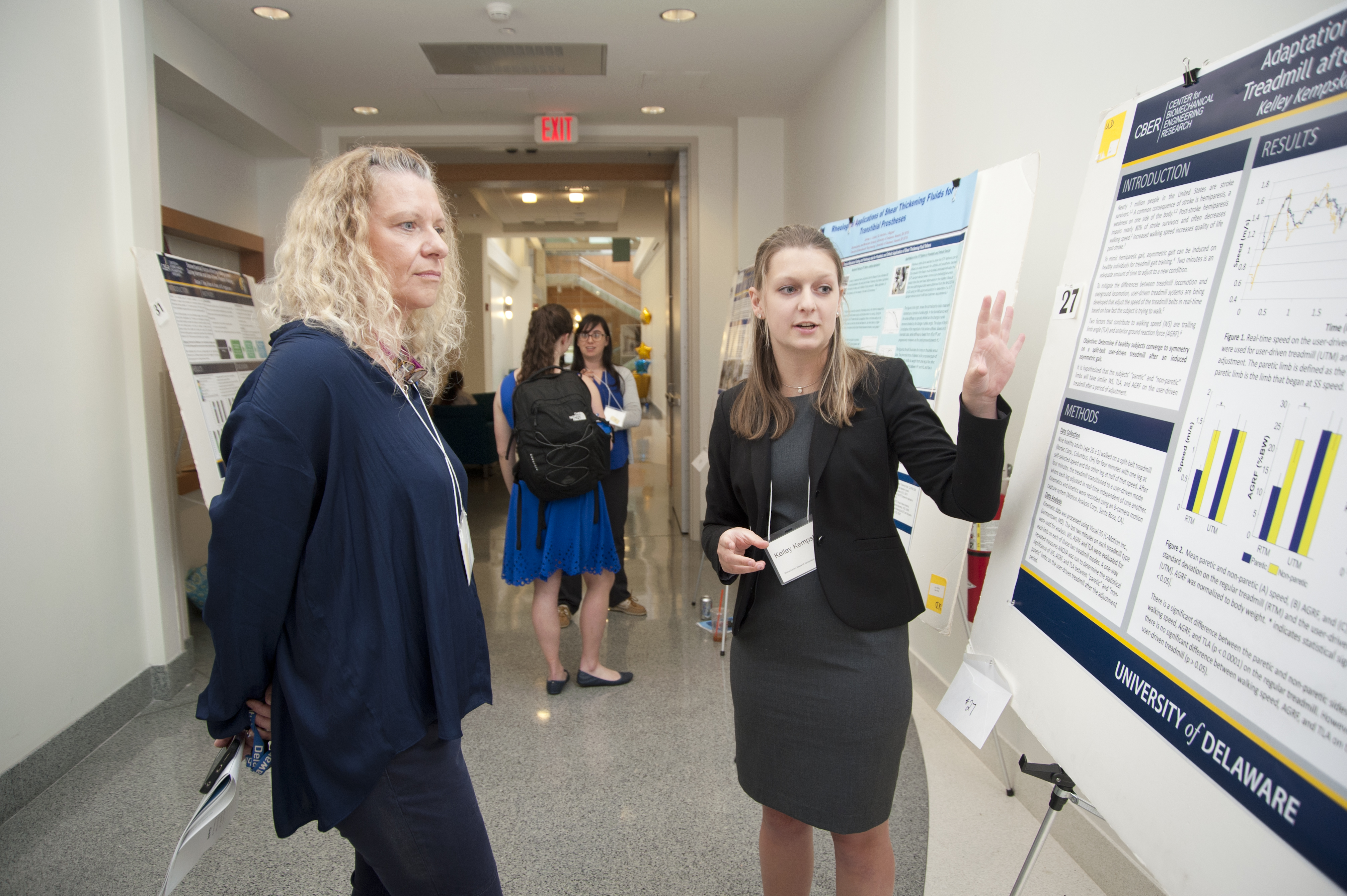
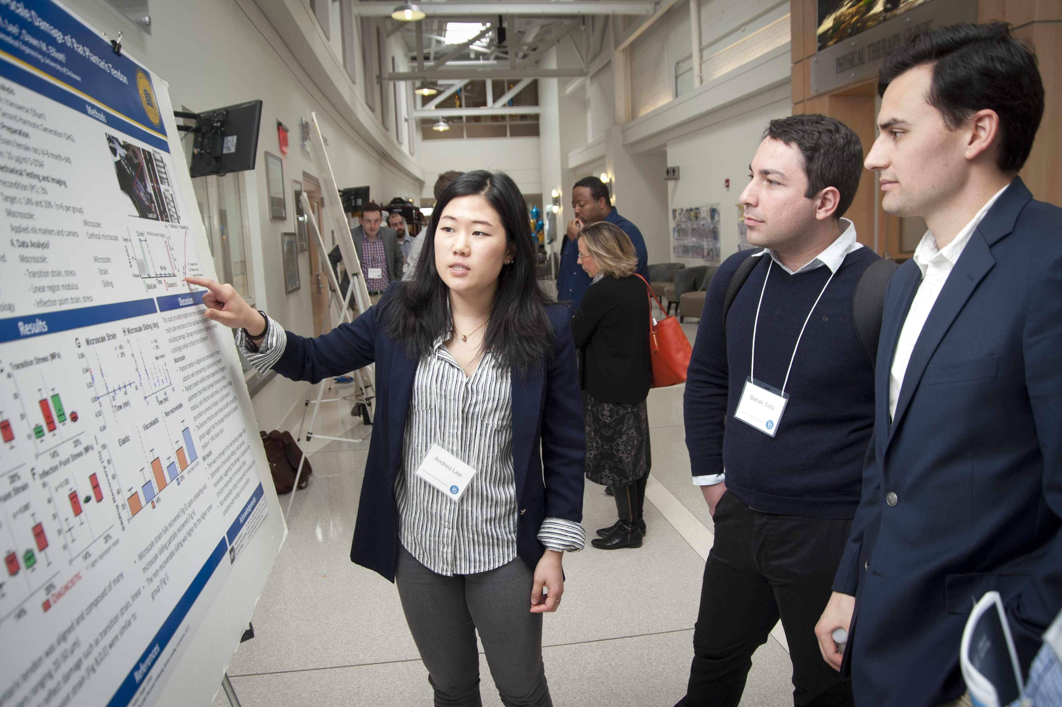
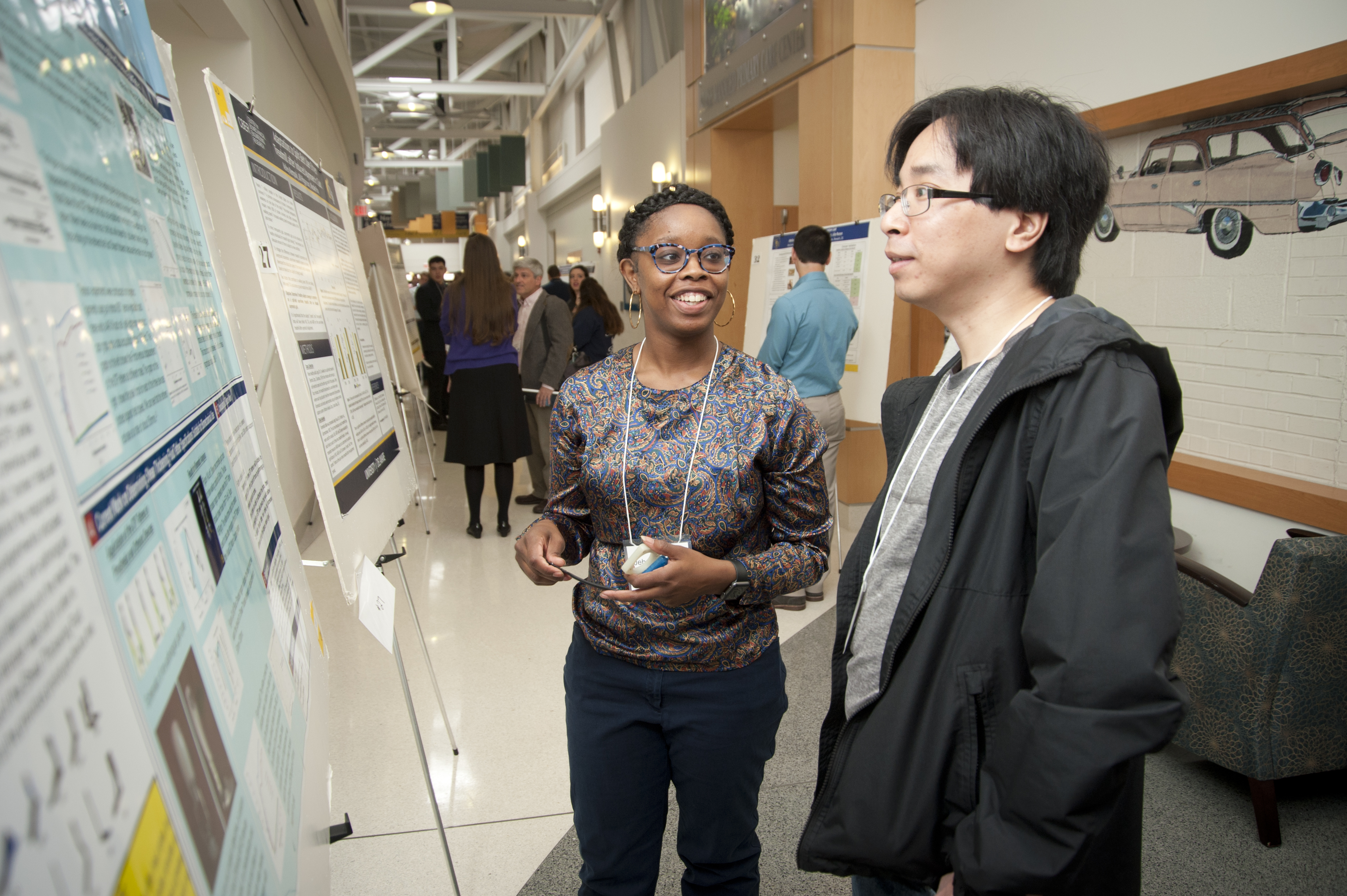
Past Event Photos


Past Event Proceedings
2023 Event
Keynote Speaker- Session 1: Hani A. Awad, Ph.D.
Title: Peritendinous Adhesions: Decoding the Roles of TGF-β and Myofibroblasts

Keynote Speaker- Session 2: Carla R. Scanzello, MD, Ph.D.
Title: Osteoarthritis as a Chronic Wound: The Role of Toll-like Receptors

2019 Event

2017 Event

2015 Event

2013 Event

2011 Event
2009 Event

2007 Event

Keynote Speaker: Dr. Steven Stanhope
2005 Event

Keynote Speaker: Dr. Steven Kautz
2021 Event
Virtual Event
Keynote Speaker Session 1: James Iatridis, PhD
Title: Biomaterial Strategies for Repair and Regeneration of the Annulus Fibrosus

Keynote Speaker Session 2: Daniel Ferris, PhD
Title: Embodiment for Bionic Prostheses and Robotic Exoskeletons

2018 Event

2016 Event
2014 Event

2012 Event

2010 Event
2008 Event

Keynote Speaker: Dr. Hugh Herr
2006 Event

Keynote Speaker: Dr. Steven Kautz
2004 Event

Keynote Speaker: David Lloyd